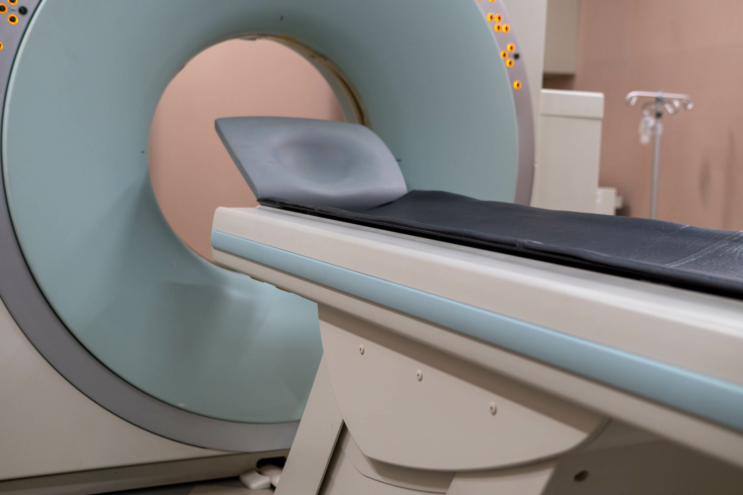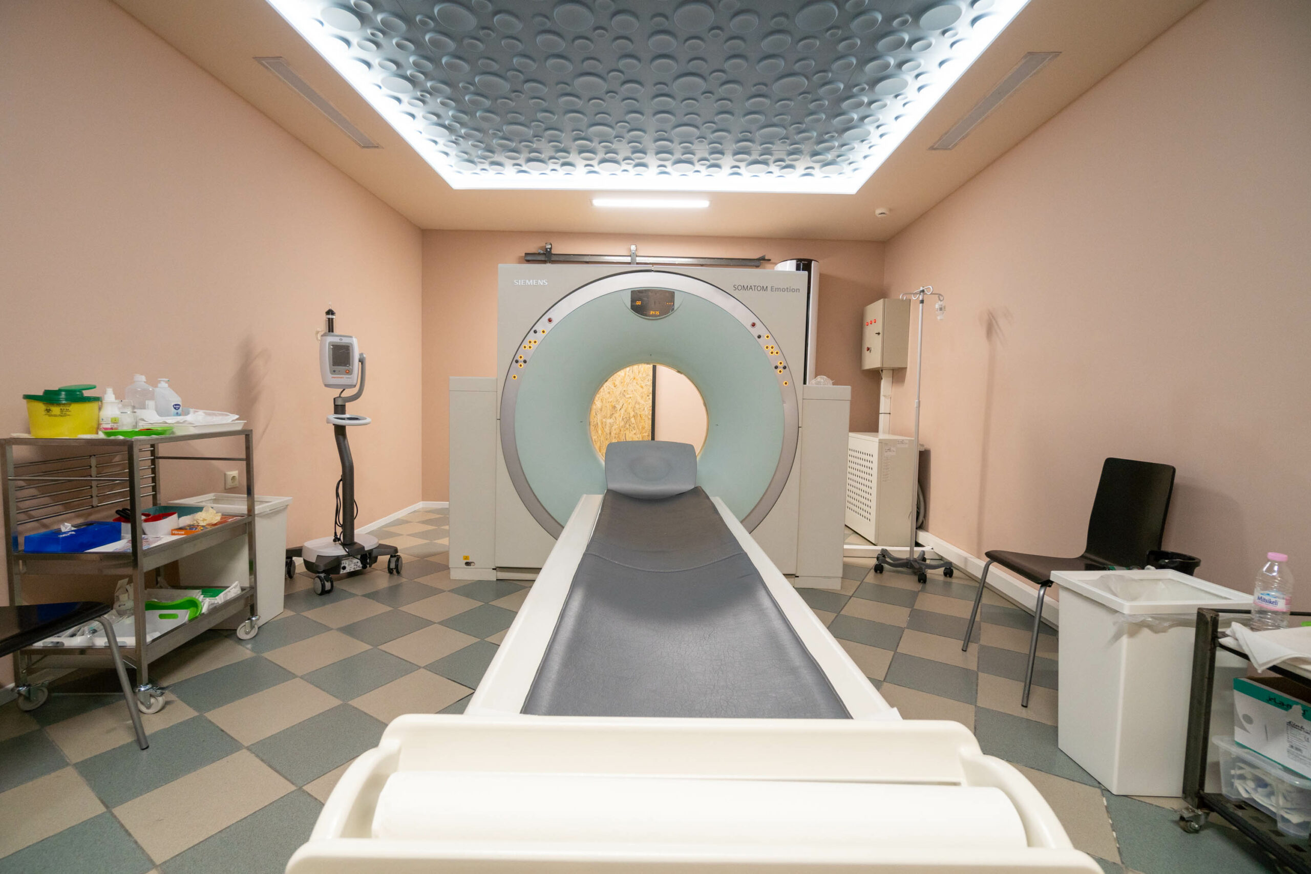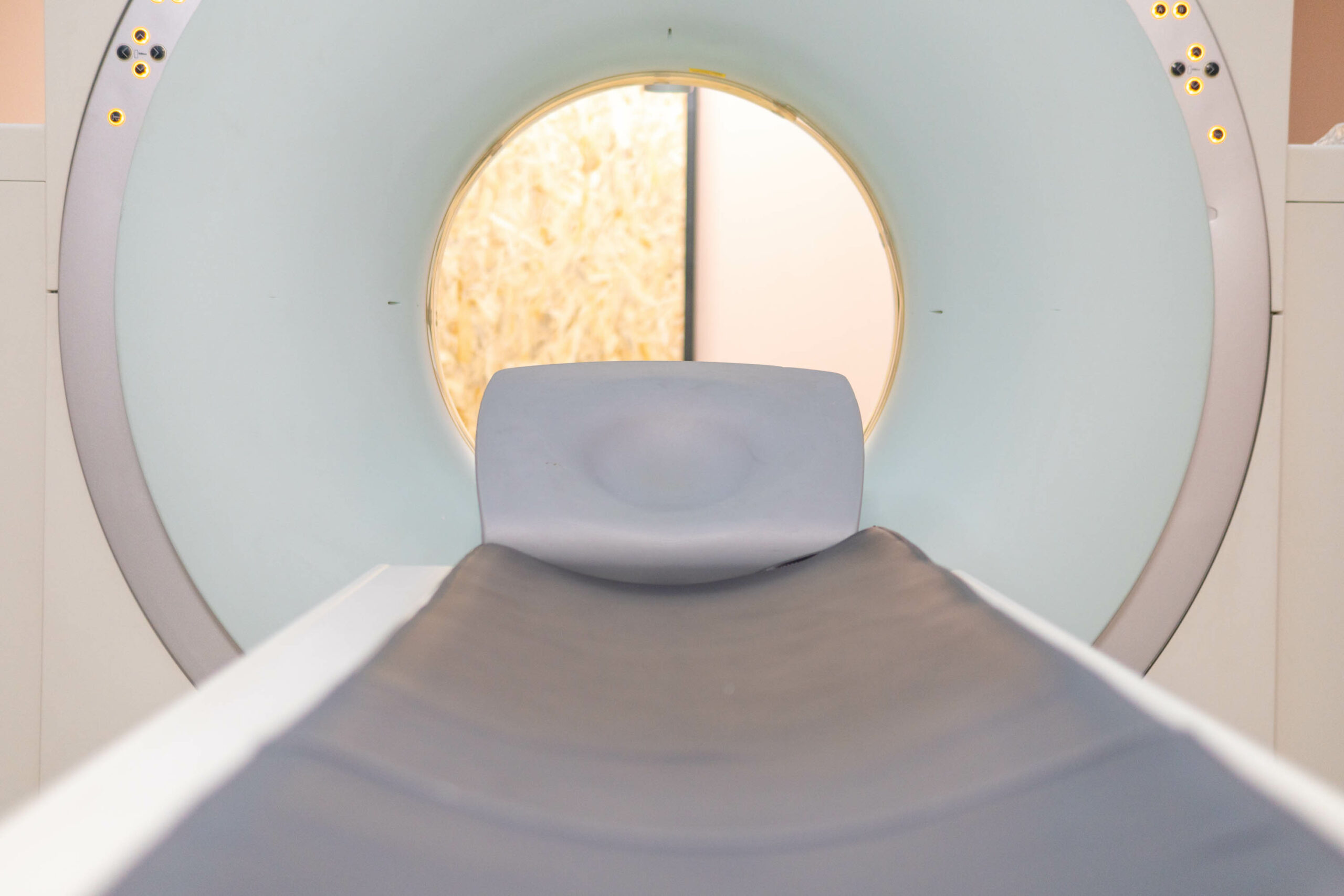
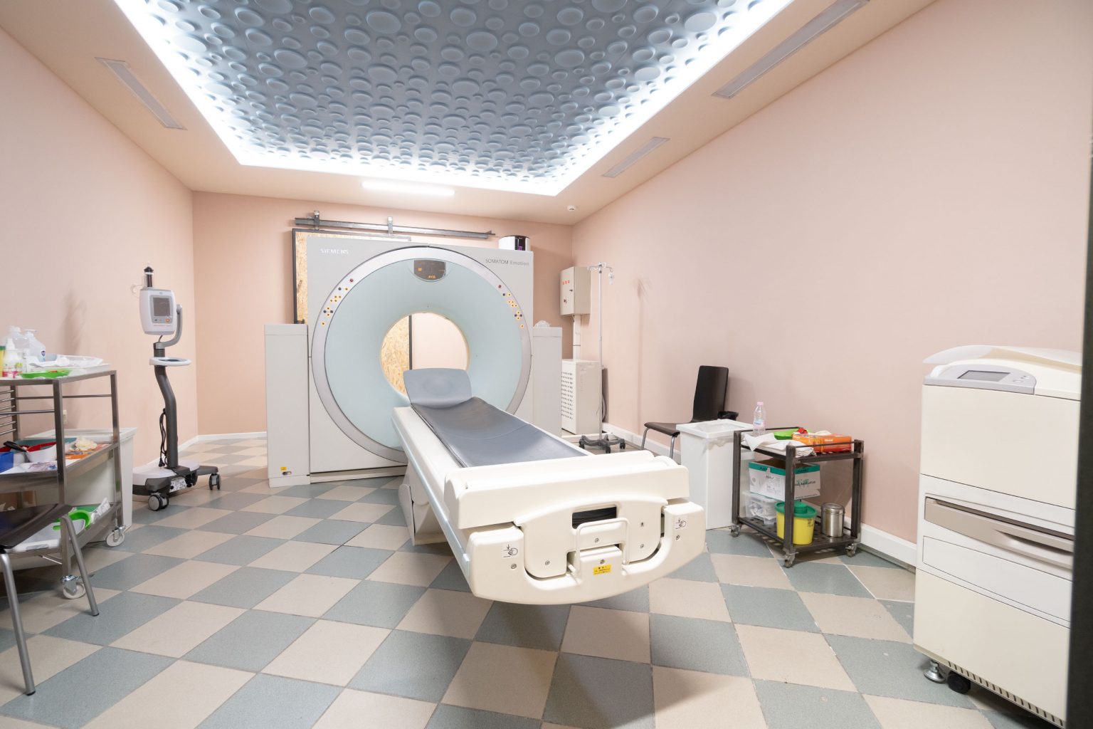
COMPUTED TOMOGRAPHY (CT SCAN)
Commonly referred to by its initials CT (Computed Tomography), this is an imaging technique that uses X-rays to scan the body in cross-sectional slices. Initially introduced in the 1970s for brain imaging, CT scans are now used to visualize all organs of the body due to their high diagnostic accuracy. With advancements such as spiral (helical) CT, it is now possible to capture images from a wider range of angles and slice depths. These images can be reconstructed even after the scan is completed, during the diagnostic phase, allowing the radiologist to create three-dimensional representations of the area under examination—something that previously only MRI (Magnetic Resonance Imaging) could achieve.
CT scans are most often performed with the simultaneous injection of an intravenous contrast agent, depending on the clinical indication and the patient’s medical history. The procedure is painless and relatively simple for the patient. In some cases (e.g., abdominal CT), the patient may be asked to fast in the morning before the scan and to ingest a special substance (such as Gastrografin) that enhances the imaging of solid organs and blood vessels. This is always done following the instructions provided by the imaging center and in coordination with the referring physician and the patient, especially regarding any known allergies.